
Case studies


Metals make up less than 0.1% of the human body by weight, yet they are essential to life, acting as cofactors for approximately one-third of all known enzymes. These ions are critical for stabilising protein structures and facilitating vital redox reactions. When metal homeostasis is disrupted, it leads to significant pathologies, including Wilson’s disease (copper accumulation), anaemia (iron deficiency), and regulated cell death pathways such as ferroptosis and cuproptosis. To study these conditions and the efficacy of metal-based drugs, researchers need to monitor the concentration and distribution of metal ions directly within the cellular environment.
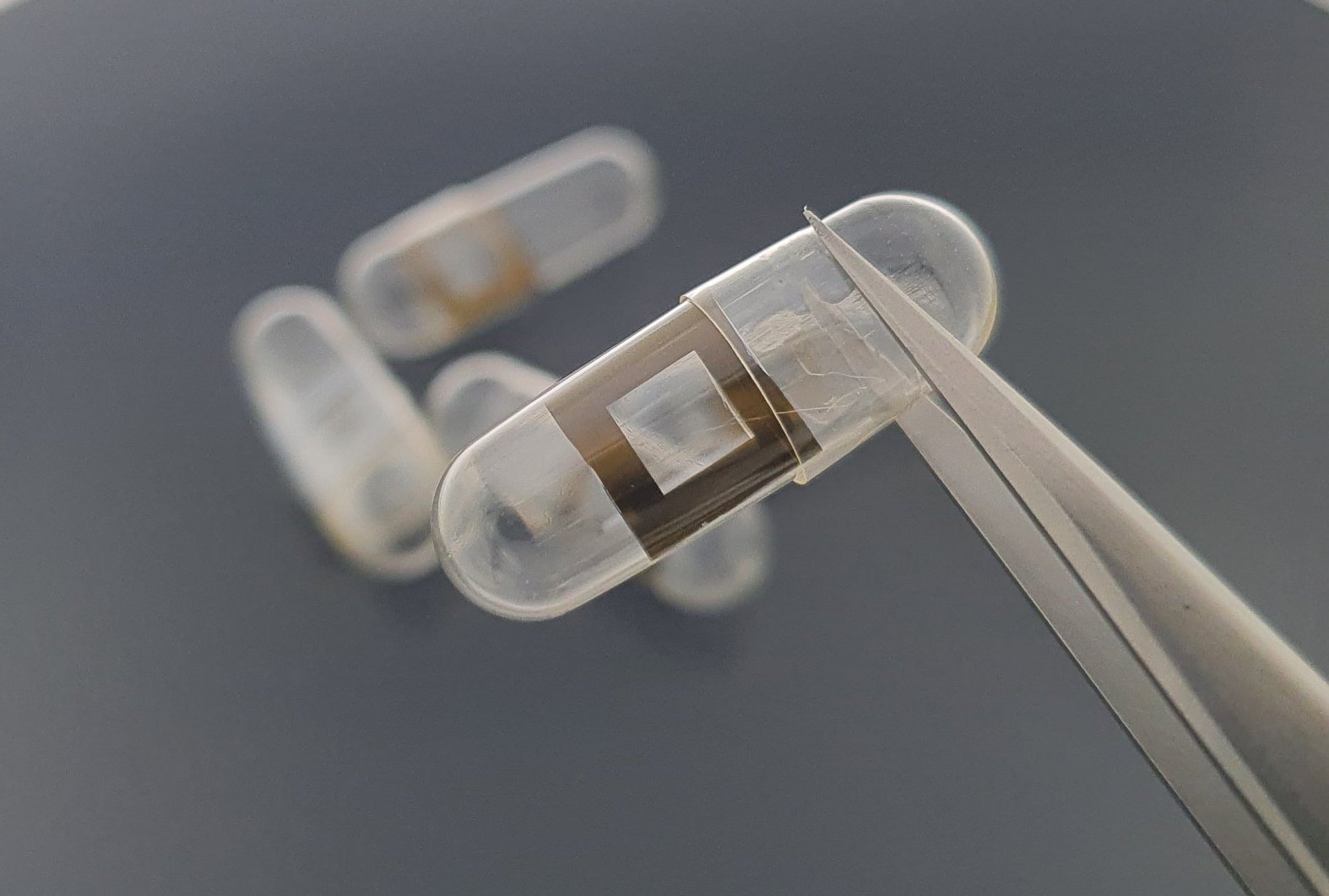
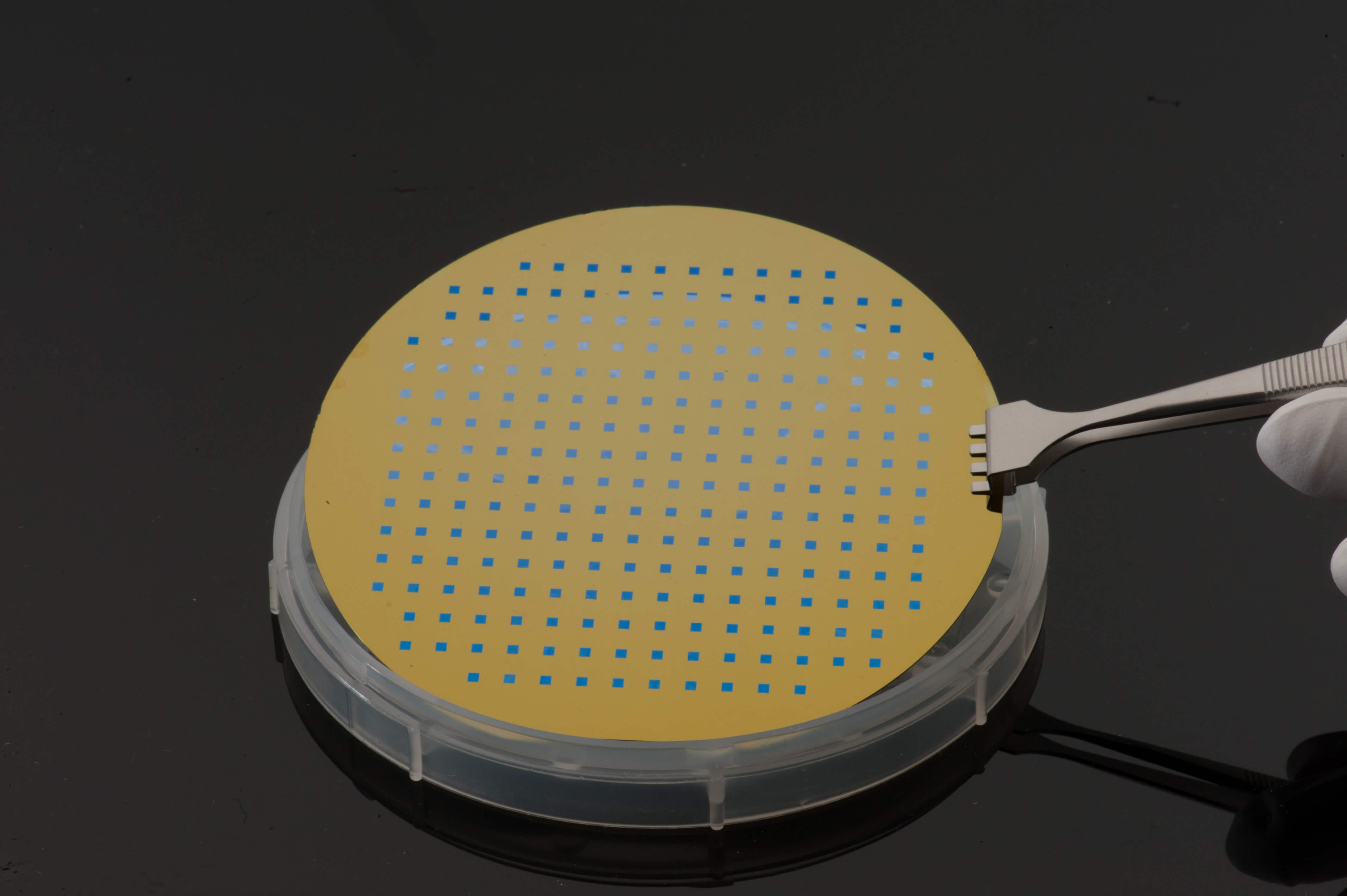
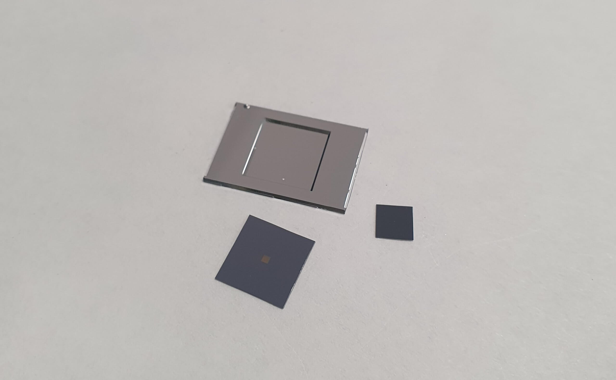
In this study, researchers utilised Silson’s 1.5 mm square SiN membranes (500 nm thick) as chemically inert substrates for culturing mammalian cell lines like MEF and HeLa. The team then established a streamlined workflow for X-ray fluorescence (XRF) and X-ray absorption spectroscopy (XAS), allowing for the submicron mapping of low-abundance metals like endogenous iron.
To maintain structural integrity of the cells and prevent the recrystallization of water, the specimens were kept at cryogenic temperatures below -130°C during all handling and transfer stages. This work demonstrates that Silson membranes are robust enough to survive cryogenic environments.
Through this work, a practical framework for investigating metal homeostasis and the mechanisms of action for new metallodrugs was established.
Read the full article, published in Chemistry Methods, here.
