
Case studies


Selenium is an essential micronutrient vital for antioxidant defense and immune function, but it operates within a very narrow therapeutic window. Understanding how different forms of selenium, such as nanoparticles and organic compounds, are processed by the body is critical for treating deficiency and potentially detoxifying heavy metals like mercury. However, identifying specific selenium metabolites in their native state within biological tissues is technically challenging due to their low concentrations and chemical sensitivity.
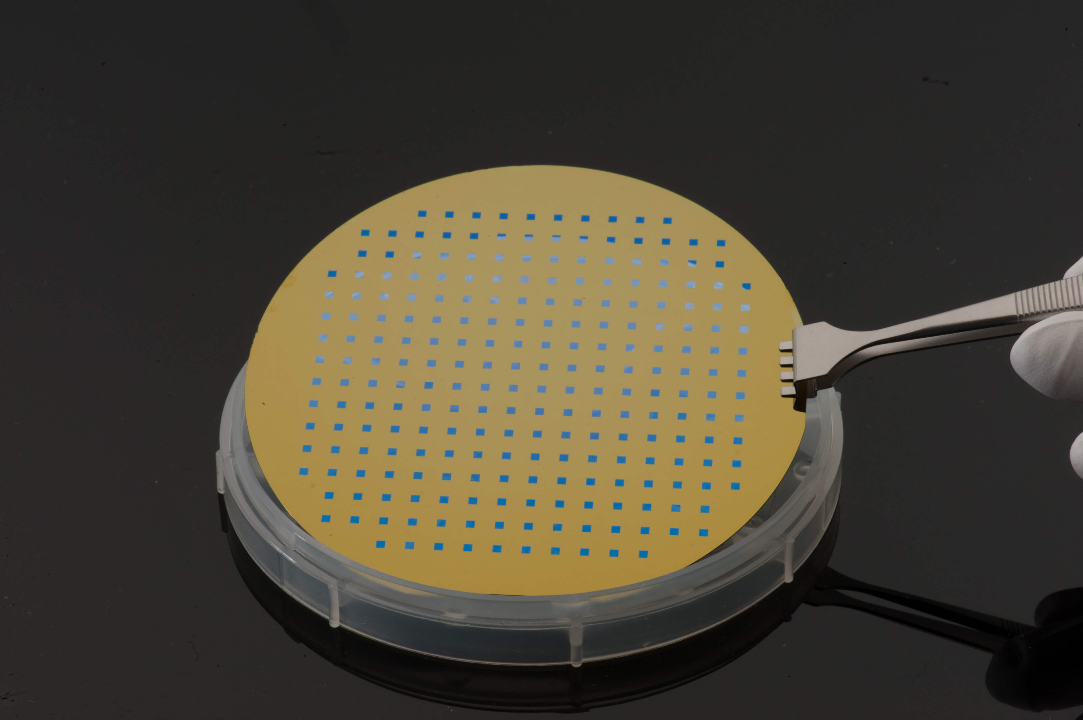
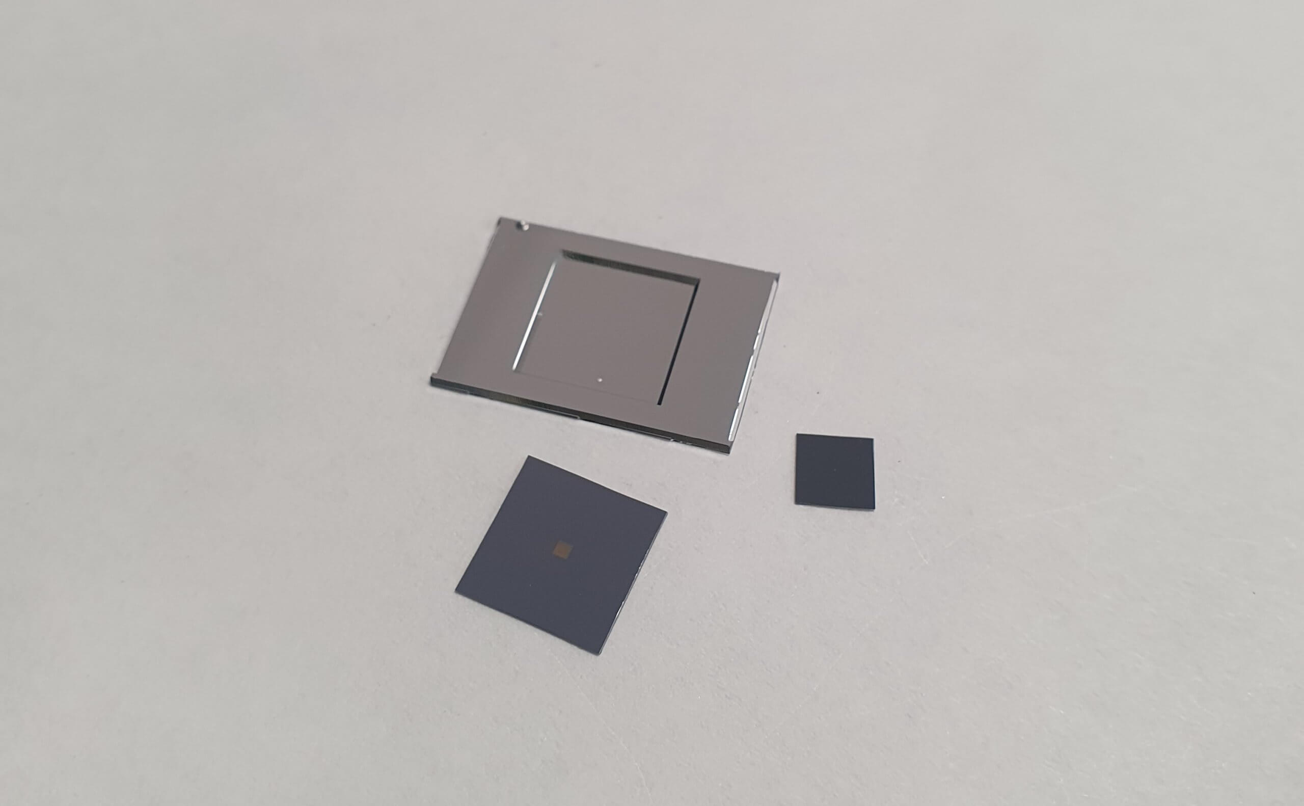
Researchers used Silson’s 500 nm thick, 5 mm square SiN windows as ultra-thin, X-ray transparent substrates to study the distribution and speciation of selenium using X-ray Fluorescence Microscopy (XFM). Tissues from mice supplemented with selenium nanoparticles and selenoneine (a compound found in marine life known to counteract methylmercury toxicity) were cryo-sectioned and mounted directly onto the SiN windows before being analyzed at the Australian Synchrotron.
This approach allowed the team to map elemental distributions with high resolution and identify that selenoneine accumulates throughout the body in its active, reduced monomer form. In contrast, selenium nanoparticles exhibited surprisingly low bioavailability when administered through food.
These results demonstrate how synchrotron X-ray techniques and high-quality substrates can reveal the complex biological fate of therapeutic compounds. This provides essential data for developing safer strategies for nutritional supplementation and utilizing selenium’s natural ability to neutralize heavy metal toxins.
Read the full article published in the Redox Report here.
